Highlighted research from Washington University chemists demonstrates innovative footprinting method to answer questions about proteins in biological systems with applications in drug design, binding, and screening.

Researchers working with Michael L. Gross, professor of chemistry in Arts & Sciences and of immunology and internal medicine at the School of Medicine, and Weikai Li, associate professor of biochemistry and molecular biophysics at the School of Medicine, have developed a novel technique for footprinting integral membrane proteins in lipsosomes, common delivery vehicles for pharmaceutical drugs, including mRNA vaccines. The new approach forges an important connection between materials science and biology and promises applications in drug design, binding, and screening. The research was published December 14 in Nature Communications and was recently selected as an editor’s highlight among papers in structural biology, biochemistry, and biophysics.
“Membrane proteins are of high importance because they participate in almost all physiological processes and represent more than 60% of drug targets,” Gross explained. “However, there are known structures of only a few hundred of these proteins compared to tens of thousands of structures for soluble proteins, which can be found in water and biological fluids. Thus, there is a large gap to be filled with new footprinting methods focused on membrane proteins.”
Footprinting uses fast chemical reactions to mark the surfaces of proteins and report on the location of amino acids. By combining footprinting with mass spectrometry, researchers can map the location of these chemical markers and observe how protein structure changes under certain conditions. Membrane proteins, as opposed to soluble proteins, have historically been challenging to mark in chemical reactions and to analyze by mass spectrometry.
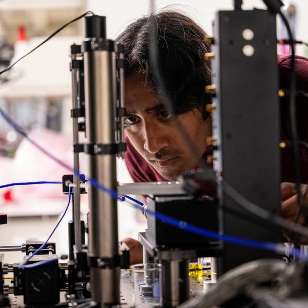
Jie Sun, lead author of the new study and a postdoctoral research associate in Gross’ laboratory, tackled this problem by attaching small nanoparticles of titanium oxide to a membrane-like material called a liposome containing a membrane protein. With laser light irradiation, she produced highly reactive free radicals in the vicinity of the liposome. Making use of the same laser beam, she conducted a fast photochemical reaction of the liposome to open it and admit the radicals to react with and mark the protein. She demonstrated that her method works for two-membrane protein systems, making it applicable to liposomes.
“A new frontier has now been opened to footprint proteins in cells instead of liposomes to answer questions about proteins in living systems,” Gross said. “This should be possible by attaching the nanoparticles to cell membranes and following our protocol. Such experiments are already underway, and we look forward to new breakthroughs in this field.”
Read the full paper, “Nanoparticles and photochemistry for native-like transmembrane protein footprinting,” in Nature Communications and related research from Sun and Gross, “Carbocation footprinting of soluble and transmembrane proteins,” in Analytical Chemistry.